High Content Screening / Automated Microscopy / Image Cytometry
Thanks to the generous support of the Medical Faculty, the Olympus scanR system has just undergone a major update/upgrade, which also includes the new AI analysis module from Olympus. More details will be posted soon.
Olympus scanR High Content Screening Station
Lighthouse has an Olympus scanR widefield microscpy system, which allows automated image acquisition and data analysis of biological samples. Other commonly used terms for this general area include image cytometry or automated microscopy. This technology is useful to "bridge the gap" between flow cytometry and microscopy.
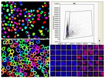 The scanR system is able to process large numbers of samples in an objective manner, enabling quantitative analysis across cells, wells, and samples. Although the total throughput is not as high as that of a flow cytometer it remains higher than that of a typical microscope, allowing entire cell populations to be examined. Unlike in flow cytometers, the samples can be examined in situ, allowing morphological information from cells and tissue sections to be collected and used for analysis. However, the images acquired can be analyzed through the use of the histograms, dot plots and even population gating, as typically used for flow cytometry.
Sample Types
Sample types which can be investigated with the scanR include both adherent and non-adherent cells and tissue sections. A climate-controlled chamber is available for living cells and time lapse experiments.
The high speed of the system means that fluorophores are much less prone to bleaching, making it possible to also use phycobiliproteins (PE, APC) for labeling your sample, in addition to the more standard microscopy fluorophores.
Sample Substrates
The scanR is very flexible with regard to what substrates can be used for the samples. However, plates and dishes with optical bottoms are recommended for the best quality images and lowest background.
-
Microtiter plates (96- or 384-well)
-
Chambered coverslips
-
Standard glass slides
-
Petri dishes
-
Standard tissue culture plates
Excitation/Emission
The scanR uses a SpectraX NIR LED Light Engine for fluorescence excitation. The possibility to use near infrared excitation/emission is unique amd makes the system particularly attractive for multiplexing fluorophores or for getting beyond regions of the spectrum where autofluorescence can be problematic.
| Excitation Wavelengths | LED | Example Fluorophores |
| 395/25 nm | Violet | DAPI, Hoechst, Brilliant Violets, other violet excitable dyes |
| 438/29 nm | Blue | ECFP, Sytox Blue |
| 475/28 nm | Cyan | EGFP, Alexa 488, FITC, CY2, (EYFP) |
|
555/28 nm *(mCh 575/25) |
Green *(Orange) |
CY3, TRITC, Alexa 546/555, PE *(mCherry, Alexa 594, Texas Red) |
| 635/22 nm | Red | CY5, Alexa 633/647, APC, (CY5.5), DRAQ5/7, SiR DNA |
| 730/40 nm | Teal | NIR / CY7, Alexa 750, Zombie NIR |
*Excitation for CY3 and mCherry is an "either/or" choice and not simultaneous.
Filter cubes, dichroics and emission filters are separate from the LED excitation system and can be used in different configurations.
Standard emission filters are listed below (others available upon request):
|
Emission Wavelengths |
Example Fluorophores |
| 433/24 nm | DAPI, Hoechst, BV421, Alexa 405 |
| 482/25 nm | eCFP, Sytox blue |
| 525/35 nm | eGFP, FITC, Alexa 488, CY2, PE |
| 600/37 nm | CY3, TRITC, Alexa 546/555 |
| 641/75 nm | mCherry, Alexa 594, Texas Red |
| 680/42 nm | CY5, Alexa 633/647, APC, DRAQ5/7 |
| 809/81 nm | NIR / CY7, Alexa 750, BV 786 |
Additional filter cubes are also available (for example CY5.5, em 716/40 nm) but must be used in combination with LED excitation.
IHC
Although for quantitation fluorescence is always the first choice, it is also possible to examine/quantify standard histologically stained/IHC specimens (brightfield) using the scanR system in transmitted light mode.
If you have any questions about possible labelling strategies, we would be happy to help you.
Access
Only those investigators who have been previously trained to use the Olympus scanR are allowed to use it. For information about reserving time on the instrument, please contact us.