Microscopy and Image Analysis - Overview
Below you can find an overview of systems located in the ZTZ building in the Lighthouse main lab, as well as in the BiMiC of the IMITATE building. Detailed information about individual system configurations can also be found in the Lighthouse calendar.
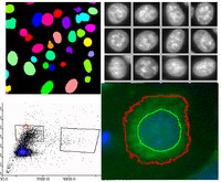
Complex Microscope Systems

Spatial Biology / Highly Multiplexed Microscopy
Lighthouse has recently installed an
Akoya Phenocycler-Fusion (CODEX) (ZTZ)
which allows the analysis of up to 100+ markers on a single slide.
More information about the Phenocycler-Fusion
 Confocal and Lightsheet Microscopy
ZTZ: Zeiss LSM 880 with AiryScan
ZTZ: Zeiss LSM 710
IMTATE: Zeiss LSM 980 NLO (multiphoton) with AiryScan
IMTATE: Zeiss CellDiscoverer 7 with LSM 900 AiryScan Scanhead
IMTATE: Zeiss Lightsheet 7
More information about the microscopes
 High Content Screening (HCS)/ Automated Microscopy
Microscopes for Image Cytometry/Automated Microscopy/HCS:
ZTZ: Olympus scanR High Content Screening Station
IMITATE: Zeiss CellDiscoverer 7 with LSM 900 AiryScan
More information about the HCS systems
Slide Scanning

Zeiss AxioScan 7 (IMITATE)
High capacity slide scanner for fluorescence or brightfield (IHC) samples. (Located in the IMITATE building)
Long-Term Live Cell Imaging

Lighthouse has a Sartorius Incucyte S3 system, which is an automated microscope contained within an incubator. It allows the independent imaging and analysis of up to 6 plates or experiments simultaneously.
More information about the Incucyte system.
Additional Widefield Systems
There are also several other microscope systems available for you to use:

Zeiss AxioImager M2m (ZTZ)
Upright widefield microscope for standard immunohistochemistry and fluorescence microscopy.
 Zeiss AxioObserver 1/
Zeiss AxioOvserver 2 (ZTZ)
Inverted fluorescence microscopes with on-stage climate chamber or heated stage, motorized XYZ stages. Tiling, Z-stacks, and more.

Zeiss Discovery.V8 (ZTZ)
Stereomicroscope. Brightfield and fluorescence (GFP/RFP).
Image Analysis
 Several workstations in the facility are reserved for offline analysis. Available software includes:
- Imaris - 3D reconstruction and analysis from Oxford Instruments
- Image Pro Plus with AutoQuant Deconvolution, 2D and 3D analysis and reconstruction
- Zeiss Zen Blue / Black
- Evident/Olympus scanR analysis, with Deep Learning AI
- Cell Profiler
- ImageJ / FiJi
- Akoya Inform Software
- QuPath
- CytoMap
Biosafety Information
Live cell imaging of samples of biosafety level S2 (Gentechnikgesetz/ Infektionsschutzgesetz) requires that you first fill out a
Lighthouse Biosafety Form
Note:
It is not necessary to fill out the checklist for every appointment. However, it should be filled out for each project, including experiments involving different cell types, treatments or sample origin.
Additional Links
General Microscopy and Image Analysis Links
Lighthouse Microscopy Calendar